Corneal Infection Projects
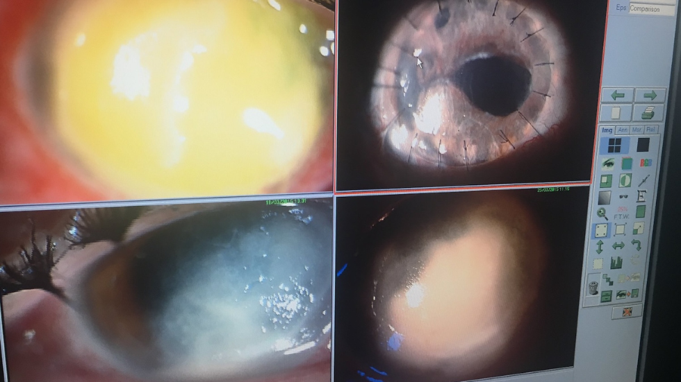
Corneal infection or microbial keratitis (MK) is a major cause of sight loss, which is associated with considerable morbidity. In temperate regions the large majority of infections are caused by bacteria. In contrast, in tropical regions fungal infections occur more frequently. We do research on the epidemiology, prevention, diagnostic tests and treatment. Currently we are working on randomised controlled trials of treatment for fungal corneal infections
Global Epidemiology of Fungal Keratitis
We recently published the first global estimate of the annual incidence of fungal corneal infections, based on a systematic review of the literature. Currently, more than one million people worldwide develop fungal keratitis each year.
The Global Incidence and Diagnosis of Fungal Keratitis
Lottie Brown, Astrid Leck, Michael Gichangi, Matthew J. Burton and David W. Denning
Lancet Infectious Diseases, 2020, In Press
Epidemiology of Corneal Infection in East Africa
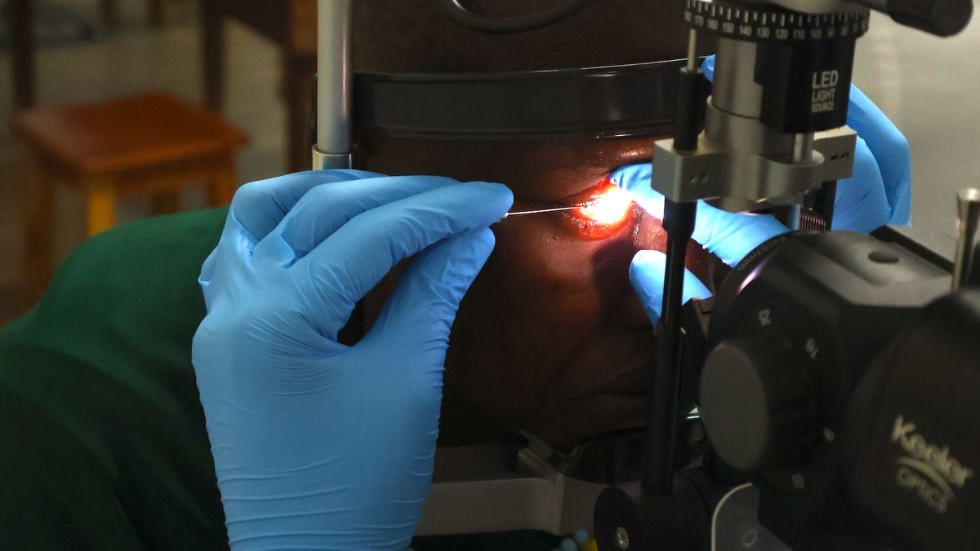
Microbial keratitis has been particularly understudied in Sub-Saharan Africa. Studies are underway to investigate the pattern and causes on MK in Tanzanian and Uganda. This involves defining the microbiology, resistance patterns and risk factors. We are also investigating the use of traditional eye medicines and the contribution these make to this disease.
Risk Factors of microbial keratitis in Uganda: a case control study
- Arunga, G. Kintoki, S. Gichuhi, J. Onyango, B. Ayebazibwe, R. Newton, A. Leck, D. Macleod, V. Hu, M. Burton
Ophthalmic Epidemiology, 2020 27(2): 98-104
Factors associated with poor presenting vision among patients with microbial keratitis in Uganda
Simon Arunga, Gladys Atto, Bosco Ayebazibwe, John Onyango, David Macleod, Victor H. Hu, Matthew J. Burton
JOECSA, 2020, In Press
The impact of microbial keratitis on quality of life in Uganda
S.Arunga, G.Wiafe, E.Habtamu, J.Onyango, S.Gichuhi, A.Leck, D.Macleod, V.H.Hu, M.J.Burton
BMJ Open Ophthalmology, 2019, 4(1):e000351
Epidemiology of Microbial Keratitis in Uganda: a cohort study
S.Arunga, G.M.Kintoki, J.Mwesigye, B.Ayebazibwe, J.Onyango, J.Bazira, R.Newton, S.Gichuhi, A.Leck, D.Macleod, V.H.Hu, M.J.Burton
Ophthalmic Epidemiology, 2020, 27(2): 121-131
The management of microbial keratitis within Uganda’s primary health system: a situational analysis
S.Arunga, N.Kyomugasho, T.Kwaga, J.Onyango, A.Leck, D.Macleod, V.H.Hu, M.J.Burton
Wellcome Open Research, 2019, 4:141
Traditional eye medicine use in microbial keratitis in Uganda: a mixed methods study
S Arunga, A Asiimwe, E Apio. O, G Kagoro-Rugunda, B Ayebazibwe, J Onyango, R Newton, A Leck, D Macleod, V H Hu, J Seeley, M J Burton
Wellcome Open Research, 2019, 4:89
Delay along the care seeking journey of patients with Microbial Keratitis in Uganda
S.Arunga, G.M.Kintoki, S.Gichuhi, J.Onyango, R.Newton, A.Leck, D.Macleod, V.H.Hu and M.J.Burton
Ophthalmic Epidemiology, 2019, 26(5):311-320
Bilateral fungal keratitis in an HIV positive patient with asymptomatic genitourinary candidiasis in Uganda.
S.Arunga, T.Kwaga, A.Leck, V.H.Hu, M.J.Burton
Medical Mycology Case Reports, 2018, 22:14-17
Microbial keratitis in East Africa: why are the outcomes so poor?
M.J.Burton, J.Pithuwa, E.Okello, I.Afwamba, J.J.Onyango, F.Oates, C.Chevallier and A.B.Hall
Ophthalmic Epidemiology, 2011; 18 (4): 158-163
Epidemiology, Pathophysiology and In Vivo Confocal Microscopy of Corneal Infection in India
Despite adequate antimicrobial therapy, severe corneal inflammation often progresses, resulting in corneal necrosis, thinning and perforation. In tropical climates, fungal infection can account for more than half of all corneal ulcers and can be difficult to manage. Although the pathophysiology of this process has not been investigated in detail, it is likely that proteases (such as matrix metallopeptidases) that are produced by the inflammatory cell infiltrate as well as the pathogenic organism may play a role in corneal destruction.
Initial diagnosis of the causative organism in MK can be a challenge. In specialised ocular microbiology laboratories, microbial culture is often only positive in up to 70% of cases. Laser scanning in vivo confocal microscopy (IVCM) offers a high-resolution method of direct visualization of many pathogenic organisms as well as the host cellular responses over time.
In this study conducted with colleagues at Aravind Eye Hospital in Tamil Nadu, South India, we investigated the diagnosis and pathophysiology of severe MK in a setting with high incidence of disease. To ascertain the genes and pathways involved in pathophysiology, transcriptome analysis was done to compare bacterial and fungal corneal ulcer tissue (excised from patients at corneal transplantation) with non-infected normal cadaver cornea. A prospective observational cohort study was conducted to follow a cohort of approximately 250 severe MK patients over a 3 week period from time of presentation in order to identify predictors of clinical progression. Corneal scrape specimens obtained from this cohort were used to evaluate gene expression changes in these patients and to validate transcriptome results. Finally, IVCM was evaluated in this cohort for its diagnostic accuracy in severe ulcers, as well as its utility in monitoring disease progression or resolution.
Cellular morphological changes detected by laser scanning in vivo confocal microscopy associated with clinical outcome in fungal keratitis.
J.Chidambaram, N.Prajna, S.Palepu, S.Lanjewar, M.Shah, S.Elakkiya, P.Lalitha, D.MacLeod, and M.J.Burton
Scientific Reports, 2019, 9(1):8334
Epidemiology, Risk Factors and Clinical Outcomes in Severe Microbial Keratitis in South India
J.D.Chidambaram, N.V.Prajna, P.Srikanthi, S.Lanjewar, M.Shah, S.Elakkiya, P.Lalitha and M.J.Burton
Ophthalmic Epidemiology, 2018, 25 (4): 297-305
In vivo confocal microscopy cellular features of bacterial, fungal and Acanthamoeba keratitis
J.D.Chidambaram, N.V.Prajna, S.Palepu, S.Lanjewar, M.Shah, S.Elakkiya, D.Macleod, P.Lalitha and M.J.Burton
American Journal of Ophthalmology, 2018, 190:24-33
Persistence of Innate Immune Pathways In Late Stage Human Bacterial And Fungal Keratitis: Results From A Comparative Transcriptome Analysis
J.D.Chidambaram, S.Kannambath, S.Palepu, M.Shah, P.Lalitha, S.Elakkiya, J.Bauer, N.V.Prajna, M.J.Holland, M.J.Burton
Frontiers in Cellular and Infection Microbiology, 2017, 7:193
In vivo confocal microscopy appearance of Fusarium and Aspergillus species in fungal keratitis
J.D.Chidambaram, N.V.Prajna, N.Larke, D.Macleod, P.Srikanthi, S.Lanjewar, M.Shah, P.Lalitha, S.Elakkiya, M.J.Burton
British Journal of Ophthalmology, 2017, 101(8):1119-1123
Prospective study of the diagnostic accuracy of the In Vivo Laser Scanning Confocal Microscope for Severe Microbial Keratitis
J.D.Chidambaram, N.V.Prajna, N.L.Larke, S.Palepu, S.Lanjewar, M.Shah, S.Elaikkya, P.Lalitha, N.Carnt, M.H.Vesaluoma, M.Mason, S.Hau and M.J.Burton
Ophthalmology, 2016, 123 (11): 2285-2293